
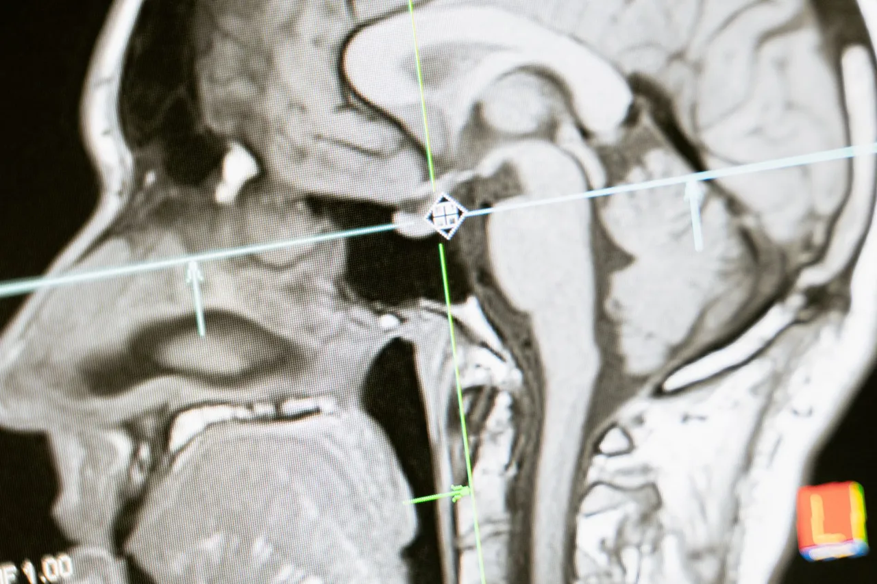
Researchers at MIT, Boston Children’s Hospital, and Inria have developed E(3)-Pose, a deep learning model designed to track fetal head movement during MRI and help produce detailed three-dimensional brain images. Presented at the 2026 European Conference on Computer Vision, the work addresses a challenge in prenatal diagnosis: movement can compromise scans used to investigate suspected brain abnormalities identified during routine ultrasound.
Fetal MRI captures two-dimensional slices that are combined into a high-resolution volume. When the head shifts, those slices can be acquired at inconsistent angles, creating coverage gaps that may conceal abnormalities or lead to incorrect diagnoses. Rather than relying entirely on training data to learn physical principles, E(3)-Pose incorporates the fetal head’s left-right symmetry directly into its neural network architecture. This geometric constraint helps the model determine the brain’s orientation. Lead author Ramya Muthukrishnan, an MIT doctoral student, pursued the project after observing the frustration of a pregnant patient and a radiologist when an uncomfortable, lengthy scan ended without a complete, high-quality image.
The approach could support earlier detection of developmental problems, while greater automation could make scans faster and more comfortable. However, the model has primarily been trained on third-trimester fetuses. Muthukrishnan hopes to adapt it for the second trimester and earlier, when smaller fetuses have more room to move within the uterus. Earlier detection can make intervention easier. Beyond fetal MRI, potential applications include imaging other difficult anatomy and tracking three-dimensional objects in autonomous driving.